Develop and apply appropriate representation and analysis techniques for digital signals and images
Hello, dear friend, you can consult us at any time if you have any questions, add WeChat: daixieit
Introduction
This Assessment Pack consists of a detailed assignment brief, guidance on what you need to prepare. You’ll also find information on this page to guide you on how, where, and when to submit. There will be a support session for this coursework on Thursday 14th Dec 2023 during the class. If you need additional support, please make a note of the services detailed in this document.
This assignment constitutes 25% of the total module assessment mark. You are expected to write a report to summarise your work on the tasks defined in the assignment brief below, particularly the Table 1 which gives a template of how your report shall be structured. Your report shall start with a very short introduction describing the image enhancement methods used, including histogram equalisation and gamma correction.
Your report has a word limit of 1000 words, excluding the references/appendices it might have.
You should use Harvard reference style in the report.
Your submission should be one zipped file which includes:
o the report in word or pdf
o the Matlab program you designed as a .m file, with comments included
o all the images used in the assignment
Your submitted zipped file should be accessible, i.e., don’t use password protection.
Teaching into assessment:
The assignment is going to be discussed at the lecture on 14/12/2023. The background of this assignment will be introduced and the tasks will be explained. Questions from students will be addressed during this support session.
Assignment brief
This assignment is designed to give you an insight into selected aspects of digital image processing which can be applied to different areas, varying from cancer diagnosis, remote sensing, motion measurement and even modern art work. You are asked to produce your image processing programs in Matlab, apply these programs and evaluate the results to obtain findings.
This assignment will enable you to:
· develop and apply appropriate representation and analysis techniques for digital signals and images
· design, analyse, select, and apply 1-D and 2-D digital filters
· create, and analyse the performance of, processing systems for digital signals and/or images for particular practical applications based on analysis of the signals/images using an appropriate fast prototyping software development platform.
The above items correspond to the Intended Learning Outcomes 2, 3 & 4 of module descriptor.
This assignment consists of three tasks (T1, T2 & T3).
The first task (T1) is to make your own histogram equalisation program, apply it to given grey level images (see Fig. 1) and evaluate the results obtained, including referring to the assumptions used in histogram equalisation and checking the statistics of the images before/after the histogram equalisation at different levels.
For Fig. 1a, it is an MRI (magnetic resonance image) frame from (Bhuvaji et al. 2000). In classical computer aided diagnosis, your image enhancement program could serve as a pre-processing step to finally classify the image into 4 different types: glioma tumour, meningioma tumour, pituitary tumour and no tumour.
Fig. 1b is from our own experiments and it is used to calibrate the resolution of an imaging system. Again your image enhancement program can be used as a step-stone to enable accurate identification of how many black-white line stripes there are within the image frame.
The second task (T2) is to apply gamma-correction to enhance the images given in Fig. 1. For each case, evaluate different setups (10x) of the gamma correction, show the histograms obtained. Unlike T1, you can use the Matlab program (imadjust) for gamma correction, i.e., you are not required to design a gamma-correction program yourself in T2.
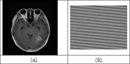
Fig. 1. Image samples: (a) Brain tumour sample from (Bhuvaji et al. 2000); (b) Black-white stripe pattern for calibrating an imaging system.
With the data obtained from T1 & T2, the third task (T3) asks you to compare the results from T1 & T2. This is an open-ended exercise but your findings shall be supported with evidence collected from T1 & T2.
For more information on histogram equalisation, see (Gonzalez & Woods 1992, Petrou & Bosdognianni 2000, Sonka et al. 1999). For gamma correction, you can refer to the help file from (Mathworks 2023).
Fig. 1a and 1b can be downloaded from EL3147’s Blackboard / Assignments folder.
References:
Bhuvaji, S., et al. (2020). Brain Tumor Classification (MRI). Available at: https://github.com/sartajbhuvaji/brain-tumor-classification-dataset (Accessed 17/11/2023).
Gonzalez, R. C. and Woods, R. E. (1992), Digital Image Processing, Addison-Wesley Publishing Company.
Petrou, M. and Bosdognianni, P. (2000), Image Processing – the Fundamentals. John Wiley & Sons Ltd.
Sonka, M., et al. (1999), Image Processing, Analysis, and Machine Vision, 2nd Edition, PWS Publishing.
Mathworks. (2023). ‘Gamma correction’, Matlab Documentation. Available at: https://uk.mathworks.com/help/images/gamma-correction.html (Accessed: 21/11/2023).
Late work
Work submitted electronically may be submitted after the deadline to the same Turnitin assignment slot and will be automatically flagged as late.
Unless an extension of the hand-in deadline date has been approved in due time, lateness penalties will be applied in accordance with University policy as follows:
(Working) Days Late by Late Penalty
1 - 5 maximum mark that can be achieved: 40%
more than 5 0% given
Marking scheme
Your report should contain the following elements which will be marked in accordance with the scheme below:
Table 1. Tasks and assigned marks
|
Tasks |
Details |
Marks |
|
T1 – histogram equalisation (HE)
|
• Histogram equalisation • Case 1 – Fig. 1a • Case 2 – Fig. 1b • Critical analysis of the results using statistics • Image level (Fig. 1a & 1b) • Selected segments (Fig. 1a & 1b) |
15 15
10 10 |
|
T2 – Gamma correction (GC) |
• Gamma correction: • Case 1 – Fig. 1a • Case 2 – Fig. 1b |
10 10 |
|
T3 - Comparison |
• Comparison the results from HE and GC • case 1 – Fig. 1a • case 2 – Fig. 1b |
10 10 |
|
|
Presentation of the report |
10 |
|
|
Total |
100 |
This assignment constitutes 25% of the total module assessment mark. You should write a report for this assignment explaining your derivations, show and discuss the results.
Feedback Guidance:
Reflecting on Feedback: how to improve.
From the feedback you receive, you should understand:
· The grade you achieved.
· The best features of your work.
· Areas you may not have fully understood.
· Areas you are doing well but could develop your understanding.
· What you can do to improve in the future - feedforward.
Use the WISER: Academic Skills Development service. WISER can review feedback and help you understand your feedback. You can also use the WISER Feedback Glossary
Next Steps:
· List the steps have you taken to respond to previous feedback.
· Summarise your achievements
· Evaluate where you need to improve here (keep handy for future work):
2024-03-03
This assignment is designed to give you an insight into selected aspects of digital image processing which can be applied to different areas